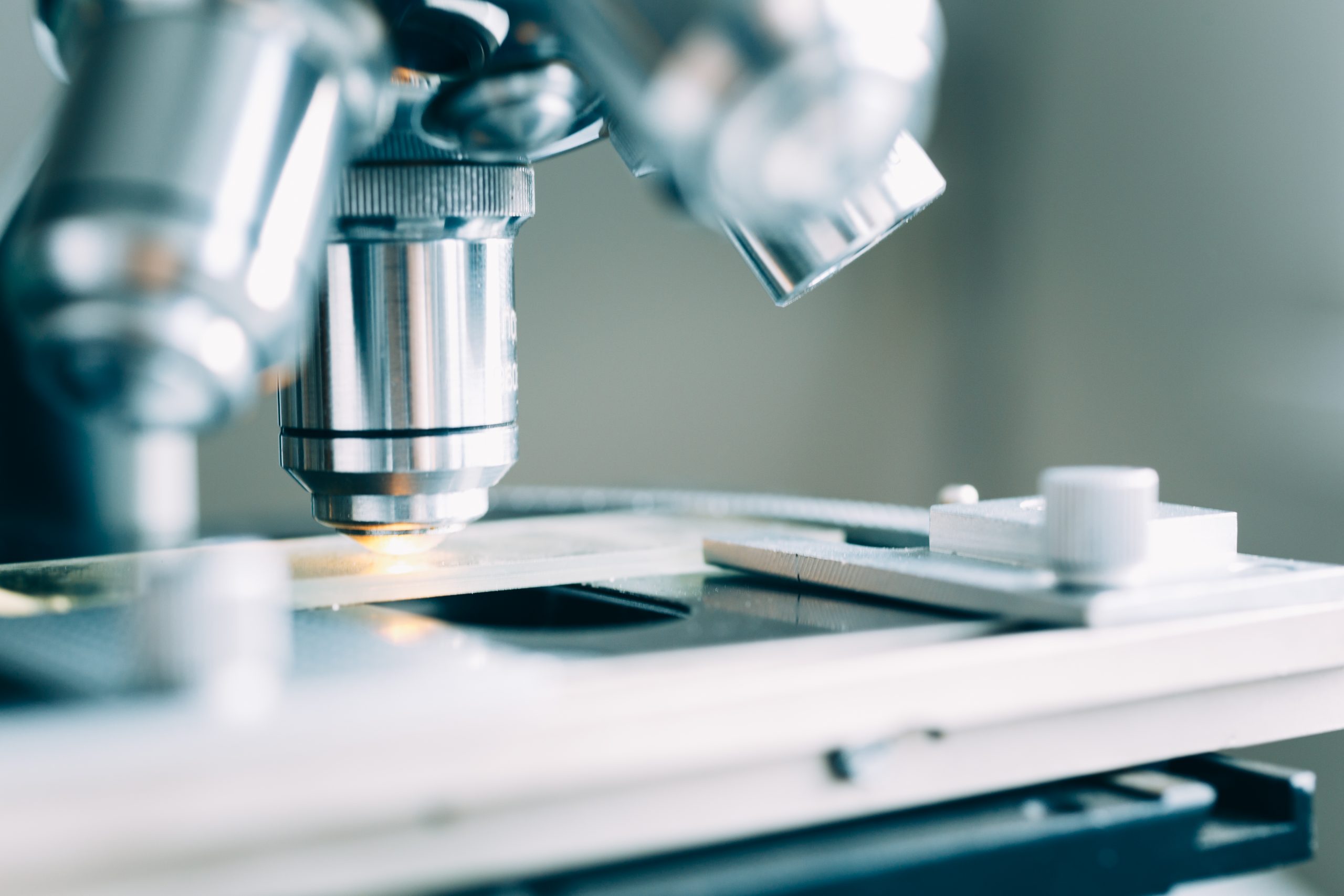
Microscopy and digital image analysis

Primary particles are vacuum dispersed in air or liquid on object-glass prior to image analysis and magnified
Optical Microscopy and digital image analysis can be used for:
- Determination of particle size distribution.
- Statistical evaluation of Particle shape, e.g. aspect ratio (ratio between breadth and length), which gives valuable extra information -complementary to laser diffraction – about particle morphology.
- Overview of particles dispersed in liquid or air.
- Observation and identification of foreign particles in the sample (pollution). Suspicion of pollution should be followed by elemental analysis by EDX/SEM or I.R./Raman to confirm foreign particles.
- Search for evidence of crystallisation in amorphous solid dispersions.
It is possible to determine the particle size distribution by number or calculated volume (0.5 µm – 2 cm). For size distribution purposes, more than 10000-50000 particles are typically processed.
For Material Experts:
Instruments and measuring principle
Particle Analytical uses an automated optical microscopy device Morphology G3 manufactured by MalvernPanalytical.
| Instrument | Morphologi G3, particle size and particle shape image analyser from Malvern |
| USP/Ph. Eur. | USP 776 /Ph. Eur 2.9.37 |
| Particle size | 0.5 µm – 2 cm |
| Dispersion pressure range | 0.5 bar – 5 bar. Dispersion pressure precision: 0.1 bar increments |
| Result | Images and diagram of particle size distribution: Size, shape, transparency, count, location. Aspect ratio, circularity, convexity, elongation, high sensitivity (H.S.) circularity, solidity fibre elongation, fibre straightness |
OR
Literature
Carlton RA (2011) Pharmaceutical Microscopy. Springer Science & Business Media.
Gamble JF, Tobyn M, Hamey R. Application of image-based particle size and shape characterisation systems in the development of small molecule pharmaceuticals. Journal of pharmaceutical sciences. 2015 May 1;104(5):1563-74.
Liang X, Xu G, Li Z, Xuan Z, Zhao H, Peng D, Gui S. Effect of micronisation on Panax Notoginseng: In vitro dissolution & in vivo bioavailability evaluations.


